I design microfluidics, computationally analyze microfluidic dynamics to better understand fluid behavior within scaffolds of porous nanofiber media, and research high-sensitivity biosensors to advance diagnostic technologies.
general Overview of Some Recent work:
Liposome Signal Amplification
As a Visiting Scholar at the Cornell Biosensors Lab, I used liposomes for signal amplification in the electrochemical detection of Cryptosporidium parvum oocysts. Species capable of generating a transducible signal (e.g. ferricyanide, sulforhodamine B) are encapsulated within a liposome tagged with nucleic acids (e.g. oligos, dendrimers), proteins (e.g. streptavidin, antigenic factors), or antibodies as recognition elements. After binding at the detection zone in the presence of the target analyte, subsequent lysis of the liposome with detergent (e.g. octyl glucoside) releases these species and produces a high degree of signal amplification.
Nanofiber Deposition Simulations
In 2013, and for the last 3 months of 2015, I developed an explicit dynamics model of nanofiber deposition during the electrospinning process, in order to overcome limitations in geometric accuracy inherent to the stochastic models used by industrial software such as GeoDict. The solver, implemented in MATLAB and C++, can simulate fiber volumes over 1,000 times larger than existing commercial solutions within just a few hours, and produces accurate anisotropy, pore size distributions, solid volume fractions, and pressure drops. I generate CFD-ready geometry from completed fiber-deposition simulations either parametrically by automating thousands of circular sweeps along fiber-defined splines with AutoLISP scripting in AutoCAD, or by exporting the data as voxels (image slices packaged in a DICOM file via an ImageJ plugin I wrote) and performing image-based meshing. I then use this geometry to create or refine a finite-volume mesh in ICEM and subsequently perform, with ANSYS Fluent, CFD analyses of dissolved-species dispersion and solution mixing in the mat, or of particulate impaction and caking.
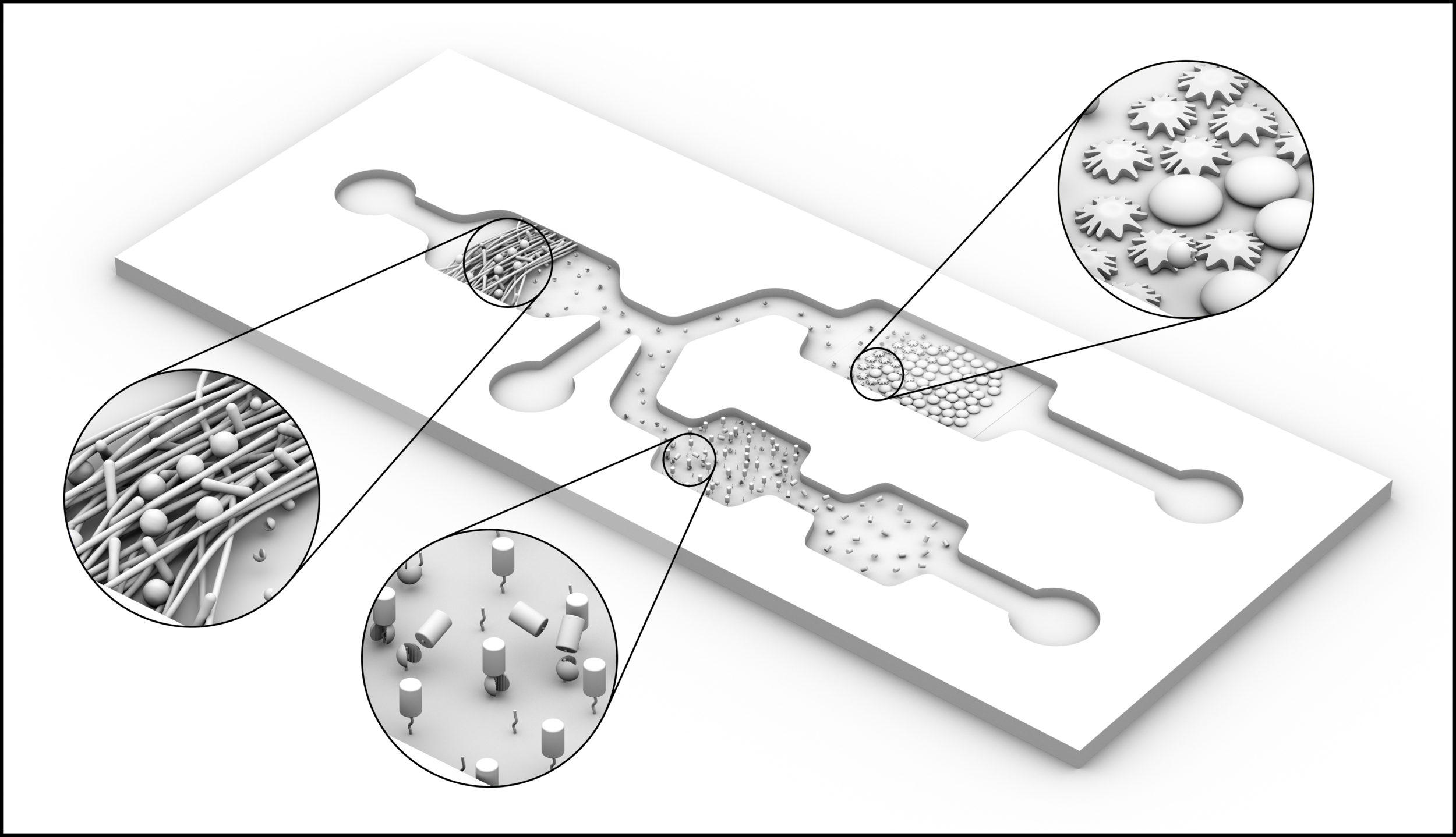
Microfluidic Platforms for Low-Cost Biosensors
PDMS is expensive and impractical for industrial use, despite the ease with which it can be used to create microchannels using an Si or metal template. At my appointments at both the Cornell University and University of Regensburg biosensor labs, I instead use PMMA (acrylic glass/Plexiglas®)—which I pattern by hot embossing with the same Si/metal templates used for PDMS soft lithography—to create microfluidic channels that are sturdier, more pressure-resistant, less absorbent, and less expensive than PDMS-based alternatives. A sample application is shown in the figure above, in which PMMA fabrication allows us to embed nanofibers into our microchannels for analyte capture (E. coli in this case), after which the captured target expresses disease/virulence factors that are captured downstream with bioreceptors capable of transducing their binding events optically (e.g. via competitive binding with a fluorophore) or electrochemically.
CAD for Machining, Micromilling, Photolithography, Molds
In 2015, I designed an easily machinable chip holder with 10 fluidic and 6 electric connections for quickly interfacing syringe-pump tubing and potentiostats to microfluidic channels. 'Nanoports' and similar single-use, glue-down connections become unfeasible when a large number of chips must be tested, and a chip holder like the one pictured permits rapid chip swapping through a zero-dead-volume connection by using PDMS ferrules (which I also cast in-house with custom molds). The chip holder is compatible with both bonded PMMA-PMMA channels (those two in the foreground) as well as with PDMS-glass channels (the casting of which is shown in the background). Lastly, the metal chip holder is compatible with harsh autoclaving/sterilization processes to remove contaminants such as the not-quite-to-scale blue bacteria pictured above.